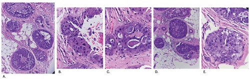
Nonmass Lesion on Breast Ultrasound
A Protean Finding
DOI:
https://doi.org/10.21141/PJP.2026.643Keywords:
nonmass lesion, breast ultrasoundAbstract
*
Downloads
References
American College of Radiology. Breast imaging reporting and data system. Accessed January 5, 2026. https://www.acr.org/Clinical-Resources/Clinical-Tools-and-Reference/Reporting-and-Data-Systems/BI-RADS
Mohan SL, Dhamija E, Gauba R. Approach to nonmass lesions on breast ultrasound. Indian J Radiol Imaging. 2024;34(4):677-87. https://pubmed.ncbi.nlm.nih.gov/39318554 https://www.ncbi.nlm.nih.gov/pmc/articles/PMC11419763 https://doi.org/10.1055/s-0044-1779589 DOI: https://doi.org/10.1055/s-0044-1779589
Choi JS, Tsunoda H, Moon WK. Nonmass lesions on breast US: an international perspective on clinical use and outcomes. J Breast Imaging. 2024;6(1):86-98. https://pubmed.ncbi.nlm.nih.gov/38243857 https://doi.org/10.1093/jbi/wbad077 DOI: https://doi.org/10.1093/jbi/wbad077
Giess CS, Chesebro Al, Chikarmane SA. Ultrasound features of mammographic developing asymmetries and correlation with histopathologic findings. AJR Am J Roentgenol. 2018;210(1):W29-38. https://pubmed.ncbi.nlm.nih.gov/29064753 https://doi.org/10.2214/AJR.17.18223 DOI: https://doi.org/10.2214/AJR.17.18223
Bahl M, Baker JA, Kinsey EN, Ghate SV. Architectural distortion on mammography: correlation with pathologic outcomes and predictors of malignancy. AJR Am J Roentgenol. 2015;205(6):1339-45. https://pubmed.ncbi.nlm.nih.gov/26587943 https://doi.org/10.2214/AJR.15.14628 DOI: https://doi.org/10.2214/AJR.15.14628
Sotome K, Yamamoto Y, Hirano A, et al. The role of contrast enhanced MRI in the diagnosis of non-mass image-forming lesions on breast ultrasonography. Breast Cancer. 2007;14(4):371-80. https://pubmed.ncbi.nlm.nih.gov/17986802 https://doi.org/10.2325/jbcs.14.371 DOI: https://doi.org/10.2325/jbcs.14.371
Yamaguchi R, Watanabe H, Mihara Y, Yamaguchi M, Tanaka M. Histopathology of non-mass-like breast lesions on ultrasound. J Med Ultrason (2001). 2023;50(3):375-80. https://pubmed.ncbi.nlm.nih.gov/36773105 https://www.ncbi.nlm.nih.gov/pmc/articles/PMC10354136 https://doi.org/10.1007/s10396-023-01286-y DOI: https://doi.org/10.1007/s10396-023-01286-y
Hong S, Li W, Gao L, et al. Diagnostic performance of elastography for breast non-mass lesions: a systematic review and meta-analysis. Eur J Radiol. 2021;144:109991. https://pubmed.ncbi.nlm.nih.gov/34638081 https://doi.org/10.1016/j.ejrad.2021.109991 DOI: https://doi.org/10.1016/j.ejrad.2021.109991
Jin ZQ, Lin MY, Hao WQ, et al. Diagnostic evaluation of ductal carcinoma in situ of the breast: ultrasonographic, mammographic and histopathologic correlations. Ultrasound Med Biol. 2015;41(1):47-55. https://pubmed.ncbi.nlm.nih.gov/25479813 https://doi.org/10.1016/j.ultrasmedbio.2014.09.023 DOI: https://doi.org/10.1016/j.ultrasmedbio.2014.09.023
Gunawardena DS, Burrows S, Taylor DB. Non-mass versus mass-like ultrasound patterns in ductal carcinoma in situ: is there an association with high-risk histology? Clin Radiol. 2020;75(2):140-7. https://pubmed.ncbi.nlm.nih.gov/31739979 https://doi.org/10.1016/j.crad.2019.10.009 DOI: https://doi.org/10.1016/j.crad.2019.10.009
Selinko VL, Middleton LP, Dempsey PJ. Role of sonography in diagnosing and staging invasive lobular carcinoma. J Clin Ultrasound. 2004;32(7):323-32. https://pubmed.ncbi.nlm.nih.gov/15293298 https://doi.org/10.1002/jcu.20052 DOI: https://doi.org/10.1002/jcu.20052
Eisenhauer EA, Therasse P, Bogaerts J, et al. New response evaluation criteria in solid tumours: revised RECIST guideline (version 1.1). Eur J Cancer. 2009;45(2):228-47. https://pubmed.ncbi.nlm.nih.gov/19097774 https://doi.org/10.1016/j.ejca.2008.10.026 DOI: https://doi.org/10.1016/j.ejca.2008.10.026
Woo J, Ryu J, Jung SM, et al. Breast radiologic complete response is associated with favorable survival outcomes after neoadjuvant chemotherapy in breast cancer, Eur J Surg Oncol. 2021;47(2):232-9. https://pubmed.ncbi.nlm.nih.gov/33213958 https://doi.org/10.1016/j.ejso.2020.08.023 DOI: https://doi.org/10.1016/j.ejso.2020.08.023
Hortobagyi GN, Connolly JL, D'Orsi CJ, et al. Breast. In: Amin MB, Edge SB, Greene FL, et al, eds. AJCC Cancer Staging Manual, 8th ed. Springer; 2017.
Harter D, O’Connor SM, Hertel JD, Calhoun BC. Pathological measurement and staging of residual breast cancer after neoadjuvant chemotherapy. Histopathology. 2023;83(3):453-64. https://pubmed.ncbi.nlm.nih.gov/37256703 https://www.ncbi.nlm.nih.gov/pmc/articles/PMC10524558 https://doi.org/10.1111/his.14966 DOI: https://doi.org/10.1111/his.14966
Ahuja S, Kiruthikasri G, Zaheer S. Evaluation of histomorphological changes in breast cancer post-neoadjuvant chemotherapy. Indian J Surg Oncol. 2024;15(2):236-40. https://pubmed.ncbi.nlm.nih.gov/38741627 https://www.ncbi.nlm.nih.gov/pmc/articles/PMC11088595 https://doi.org/10.1007/s13193-024-01876-3 DOI: https://doi.org/10.1007/s13193-024-01876-3
Downloads
Published
How to Cite
Issue
Section
License
Copyright (c) 2026 Ma. Theresa Buenaflor, David Elijah Saguil

This work is licensed under a Creative Commons Attribution-NonCommercial-ShareAlike 4.0 International License.


 Educational mini-series for authors, reviewers, pathology residents, and researchers dedicated to improving scientific writing in pathology and laboratory medicine.


 @philippinepathologyjournal