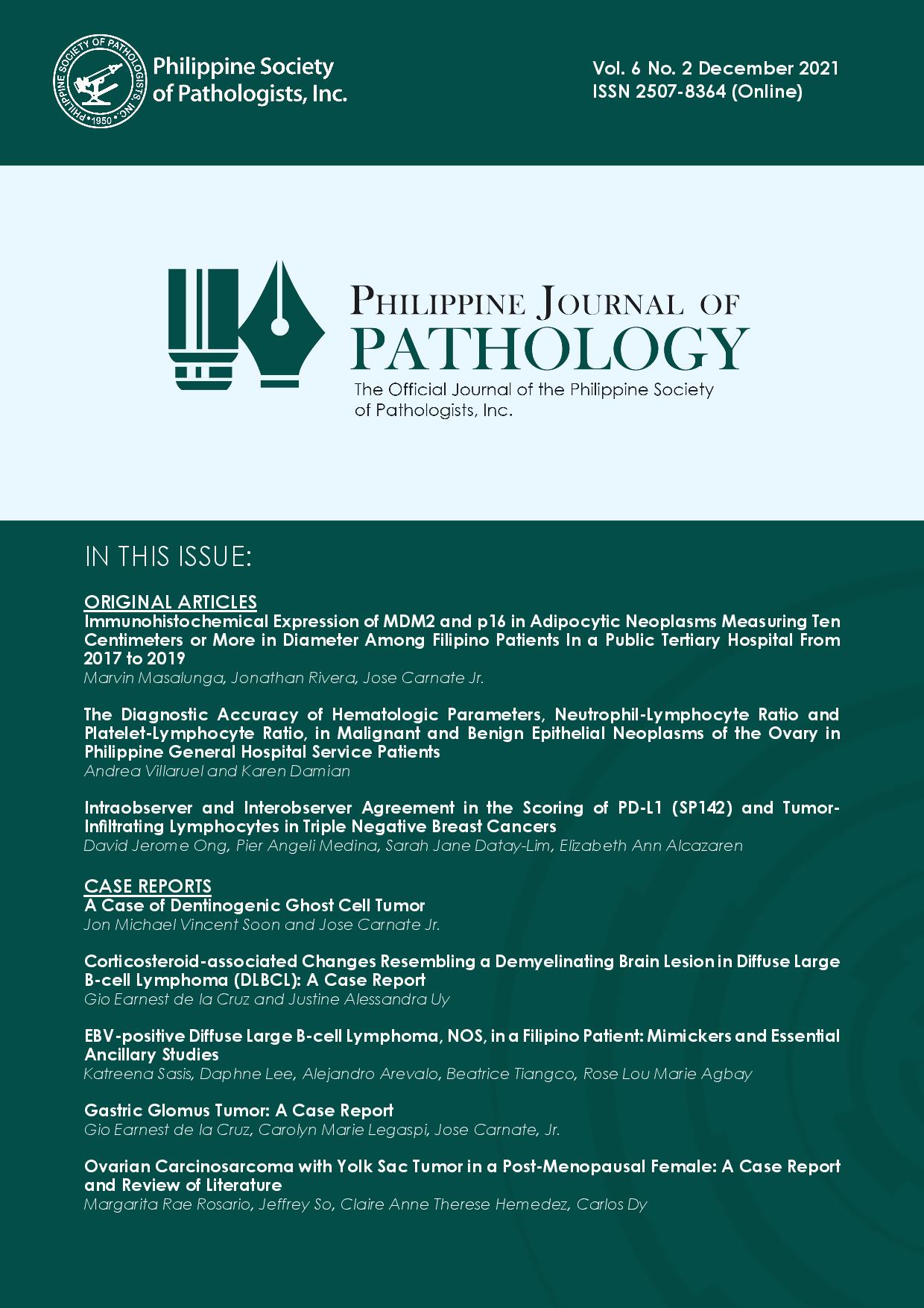
Immunohistochemical Expression of MDM2 and p16 in Adipocytic Neoplasms Measuring Ten Centimeters or more in Diameter among Filipino Patients in a Public Tertiary Hospital from 2017 to 2019
DOI:
https://doi.org/10.21141/PJP.2021.11Keywords:
neoplasms, adipose tissue, lipoma, liposarcoma, extremitiesAbstract
Introduction: A size of more than 10 cm suggests that a soft tissue tumor might be malignant. Pertinent ancillary diagnostic testing, such as immunohistochemistry (IHC) and fluorescence in situ hybridization (FISH), may be done to confirm the diagnosis. Several studies have shown that size may be a useful criterion in determining which tumors are candidates for further molecular testing. MDM2 and p16 are IHC markers for atypical lipomatous tumor/well-differentiated liposarcoma (ALT/WDLPS).
Objectives: The primary objective of this study is to determine the proportion of tumors signed out as “lipomas” from 2017 to 2019, and measuring at least 10 cm, that express MDM2 and p16 on IHC and warrant revision as ALT/WDLPS.
Methodology: This is a descriptive, retrospective cohort study in which all lipomas from 2017 to 2019 that measured at least 10 cm were included. The size, age of the patient, and location of each tumor were documented. The slides of all eligible cases were reviewed and immunohistochemically stained for MDM2 and p16. For each case, the intensity and immunoreactivity of each stain were assessed using a modified, four-tier scoring system. Fisher’s exact test was used to determine if a significant number of tumors expressed MDM2 or p16.
Results: Thirty (30) cases satisfied the inclusion and exclusion criteria. The average size of these tumors is 15.10 cm. There is no sex predilection. The most common location of these tumors is the extremities. None of the tumors expressed MDM2, and only one case was p16-positive. The case positive for p16 also showed cytologic atypia and variability in cell size, resulting in the revision of its diagnosis from lipoma to atypical lipomatous tumor. The rate of diagnosis revision after slide review and IHC studies is 3.33%.
Conclusion: None of the adipocytic tumors that measured at least 10 cm in diameter and were signed out as lipomas was MDM2 positive, and only one case was p16-positive. Thus, morphology remains the cornerstone in the diagnosis of adipocytic tumors. Careful microscopic evaluation is necessary to establish the diagnosis of malignancy in these tumors. Ancillary tests should only be considered in cases where the pathologic features are equivocal.
Downloads
References
2. Goldblum J. Soft tissues. In Goldblum J, Lamps L, McKenney J, Myers J. Rosai and Ackerman's Surgical Pathology. Philadelphia: Elsevier; 2018.
3. Billings SD, Din NU. Spindle cell lipoma and pleomorphic lipoma. International Agency for Research on Cancer; 2020. Available from: https://tumourclassification.iarc.who.int/chapter content/33/10.
4. Horvai A. Bones, joints, and soft tissue tumors. In: Kumar V, Abbas A, Aster J, editors. Robbins and Cotran pathologic basis of disease. Philadelphia: Elsevier; 2015.
5. Sbaraglia M, Tos APD, Pedeutour F. Atypical lipomatous tumour/well-differentiated liposarcoma. International Agency for Research on Cancer; 2020. Available from: https://tumourclassification.iarc.who.int/chaptercontent/33/12.
6. Tos AD, Pedeutor F. Atypical lipomatous tumor. In: WHO classification of tumours of soft tissue and bone. Lyon: International Agency for Research on Cancer; 2013.
7. Clay MR, Martinez AP, Weiss SW, Edgar MA. MDM2 amplification in problematic lipomatous tumors: analysis of FISH testing criteria. Am J Surg Pathol. 2015; 39(10):1433-9. https://pubmed.ncbi.nlm.nih.gov/26146760. https://doi.org/10.1097/PAS.0000000000000468.
8. Johnson CJ, Pynsent PB, Grimer RJ. Clinical features of soft tissue sarcomas. Ann Roy Coll Surg. 2001;83(3):203-5. https://pubmed.ncbi.nlm.nih.gov/11432141. https://www.ncbi.nlm.nih.gov/pmc/articles/2503577.
9. National Institute for Health and Clinical Excellence. Guidance on cancer services: improving outcomes for people with sarcoma. London: National Institute for Health and Clinical Excellence; 2006.
10. National Institute for Health and Care Excellence. Sarcoma quality standard. NHS England; c2015. Available from: https://www.nice.org.uk/guidance/qs78/resources/sarcoma-2098854826693.
11. Casali P, Blay JY, ESMO/CONTICANET/EUROBONET Consensus Panel of experts. Soft tissue sarcomas: ESMO clinical practice guidelines for diagnosis, treatment and follow-up. Ann Oncol. 2010;21(5):198–203. https://pubmed.ncbi.nlm.nih.gov/20555081. https://doi.org/10.1093/annonc/mdq209.
12. Okada K. Points to notice during the diagnosis of soft tissue tumors according to the “clinical practice guideline on the diagnosis and treatment of soft tissue tumors.” J Orthop Sc. 2016;21(6):705-12. https://pubmed.ncbi.nlm.nih.gov/27471013. https://doi.org/10.1016/j.jos.2016.06.012.
13. National Health Service. Ultrasound screening of soft tissue masses in the trunk and extremity. London: NHS Bristol NHS Trust; 2016.
14. Mahendra B. Challenges in the diagnosis of lipomatous tumours. J Diagn Pathol. 2014; 9(1):1-17. http://doi.org/10.4038/jdp.v9i1.7640.
15. Clay MR, Martinez AP, Weiss SW, Edgar MA. MDM2 andCDK4 immunohistochemistry: should it be used in problematic differentiated lipomatous tumors? A new perspective. Am J Surg Pathol. 2016; 40(12):1647-52. https://pubmed.ncbi.nlm.nih.gov/27508976. https://doi.org/10.1097/PAS.0000000000000713.
16. Weaver J, Rao P, Goldblum JR, et al. Can MDM2 analytical tests performed on core needle biopsy be relied upon to diagnose well-differentiated liposarcoma? Mod Pathol. 2010; 23(10):1301-6. https://pubmed.ncbi.nlm.nih.gov/20495536. https://doi.org/10.1038/modpathol.2010.106.
17. Wang S, Chan LWM, Tang X, et al. A weighted scoring system to differentiate malignant liposarcomas from benign lipomas. J Orthop Surg (Hong Kong). 2016; 24(2):216-21. https://pubmed.ncbi.nlm.nih.gov/27574266. https://doi.org/10.1177/1602400219.
18. Altun E, Yükse S, Kaygusuz G, Yıldız HY. Diagnostic importance of clinicopathologic features and P16, CD34, MDM2 expression in differential diagnosis of adipocytic tumors. Acta Orthop Traumatol Turc. 2020;54(1):59-65. https://pubmed.ncbi.nlm.nih.gov/32175898. https://www.ncbi.nlm.nih.gov/pmc/articles/PMC7243699. https://doi.org/10.5152/j.aott.2020.01.445.
19. Laurini JA, Cooper K, Fletcher CD, et al. Protocol for the examination of resection specimens from patients with soft tissue tumors. Northfield: College of American Pathologists; 2020.
20. Thway K, Flora R, Shah C, Olmos D, Fisher C. Diagnostic utility of P16, CDK4, and MDM2 as an immunohistochemical panel in distinguishing well-differentiated and dedifferentiated liposarcomas from other adipocytic tumors. Am J Surg Pathol. 2012. 36:462-69. https://pubmed.ncbi.nlm.nih.gov/22301498. https://doi.org/10.1097/PAS.0b013e3182417330.
21. Thway K. Well-differentiated liposarcoma and dedifferentiated liposarcoma: an updated review. Semin Diagn Pathol. 2019; 36(2):112-21. https://pubmed.ncbi.nlm.nih.gov/30852045. https://doi.org/10.1053/j.semdp.2019.02.006.
22. Thway K, Wang J, Swansbury J, Min T, Fisher C. Fluorescence in situ hybridization for MDM2 amplification as a routine ancillary diagnostic tool for suspected well-differentiated and dedifferentiated liposarcomas: experience at a tertiary center. Sarcoma. 2015; 2015:812089. https://pubmed.ncbi.nlm.nih.gov/25810689. https://www.ncbi.nlm.nih.gov/pmc/articles/PMC4355609. https://doi.org/10.1155/2015/812089.
23. Lee SH, Park HS, Im S, Cui Y, Kim SY, Yoo C, Yoo J. Application of previously reported lipo-related markers in atypical lipomatous tumor/well differentiated liposarcoma (ALT/WDLS) and lipoma. Int J Clin Exp Med. 2018; 11(9):9178-88. http://www.ijcem.com/files/ijcem0074439.pdf.
24. Amin M, Edge S, Greene F, et al. AJCC cancer staging manual. 8th ed. New York City: Springer; 2017.
25. Briski LM, Jorns JM. Primary breast atypical lipomatous tumor/well-differentiated liposarcoma and dedifferentiated liposarcoma. Arch Pathol Lab Med; 2018. 142(2):268-74. https://pubmed.ncbi.nlm.nih.gov/29372852. https://doi.org/10.5858/arpa.2016-0380-RSR2.
26. Al-Ani Z, Fernando M, Wilkinson V, Kotnis N. The management of deep-seated, lowgrade lipomatous lesions. Brit J Radiol. 2018; 91(1086):20170725. https://pubmed.ncbi.nlm.nih.gov/29303371. https://doi.org/10.1259/bjr.20170725.
27. Muratori F, Roselli G, Bettini L, et al. Atypical lipomatous tumor: retrospective analysis, prognostic factors and MRI in differential diagnosis. Clin Cases Miner Bone Metab. 2018; 15(2):288-93. https://arpi.unipi.it/handle/11568/948689#.YSHejMdBztQ.
28. Zhang H, Erickson-Johnson M, Wang X, et al. Molecular testing for lipomatous tumors: critical analysis and test recommendations based on the analysis of 405 extremity-based tumors. Am J Surg Pathol. 2010; 34(9):1304-11. https://pubmed.ncbi.nlm.nih.gov/20679883. https://doi.org/10.1097/PAS.0b013e3181e92d0b.
29. Creytens D. What’s new in adipocytic neoplasia? Virchows Archiv. 2019;476(1):29-39. https://pubmed.ncbi.nlm.nih.gov/31501988. https://doi.org/10.1007/s00428-019-02652-3.
30. Creytens D, Ferdinande L, van Gorp J, Van Dorpe J, Flucke U. Atypical spindle cell lipomatous tumor with benign heterologous (metaplastic) cartilaginous differentiation. Int J Surg Pathol. 2019; 27(5):521-3. https://pubmed.ncbi.nlm.nih.gov/30370805. https://doi.org/10.1177/1066896918809189.
31. Binh MBN, Sastre-Garau X, Guillou L, et al. MDM2 and CDK4 immunostainings are useful adjuncts in diagnosing well-differentiated and dedifferentiated liposarcoma subtypes: a comparative analysis of 559 soft tissue neoplasms with genetic data. Am J Surg Pathol. 2005; 29(10):1340-7. https://pubmed.ncbi.nlm.nih.gov/16160477. https://doi.org/10.1097/01.pas.0000170343.09562.39.
32. Baker SJ, Reddy EP. CDK4: a key player in the cell cycle, development, and cancer. Genes and Cancer. 2013; 3(11-12):658-69. https://pubmed.ncbi.nlm.nih.gov/23634254. https://www.ncbi.nlm.nih.gov/pmc/articles/PMC3636745. https://doi.org/10.1177/1947601913478972.
33. Yao X, Ghert M, Dickson BD, et al. An evidence-based guideline on the application of molecular testing in the diagnosis, prediction of prognosis, and selection of therapy in non-GIST soft tissue sarcomas. Cancer Treat Rev. 2020; 85:101987. https://pubmed.ncbi.nlm.nih.gov/32092619. https://doi.org/10.1016/j.ctrv.2020.101987.
34. Stojanov IJ, Mariño-Enriquez A, Bahri N, Jo VY, Woo S. Lipomas of the oral cavity: utility of MDM2 and CDK4 in avoiding overdiagnosis as atypical lipomatous tumor. Head Neck Pathol. 2019;13(2):169-76. https://pubmed.ncbi.nlm.nih.gov/29748845. https://www.ncbi.nlm.nih.gov/pmc/articles/PMC6513928. https://doi.org/10.1007/s12105-018-0928-0.
35. He M, Aisner S, Benevenia J, Patterson F, Aviv H, Hameed M. p16 immunohistochemistry as an alternative marker to distinguish atypical lipomatous tumor from deep-seated lipoma. Appl Immunohisto M M. 2009;17(1):51-6. https://pubmed.ncbi.nlm.nih.gov/18779733. https://doi.org10.1097/PAI.0b013e3181719223.
36. Kang Y, Horvai AE. p16 immunohistochemistry is less useful than MDM2 and CDK4 to distinguish dedifferentiated liposarcomas from other retroperitoneal mimics. Appl Immunohistochem Mol Morphol. 2017;25(1):58-63. https://pubmed.ncbi.nlm.nih.gov/26509911. https://doi.org/10.1097/PAI.0000000000000270.
37. Kammerer-Jacquet SF, Thierry S, Cabillic F, et al. Differential diagnosis of atypical lipomatous tumor/well-differentiated liposarcoma and dedifferentiated liposarcoma: utility of p16 in combination with MDM2 and CDK4 immunohistochemistry. Hum Pathol. 2017; 59:34-40. https://pubmed.ncbi.nlm.nih.gov/27597521. https://doi.org/10.1016/j.humpath.2016.08.009.
38. Ng W, Messiou C, Smith M, Thway K. p16 expression in fat necrosis: a potential diagnostic pitfall in the evaluation of differentiated adipocytic neoplasms. Int J Surg Pathol. 2015; 23(7):544-48. https://pubmed.ncbi.nlm.nih.gov/26173427. https://doi.org/10.1177/1066896915595465.
39. Presman B, Jauffred SF, Kornø MR, Petersen MM. Low recurrence rate and risk of distant metastases following marginal surgery of intramuscular lipoma and atypical lipomatous tumors of the extremities and trunk wall. Med Prin Pract. 2020;29(3):203-10. https://pubmed.ncbi.nlm.nih.gov/31550705. https://www.ncbi.nlm.nih.gov/pmc/articles/PMC7315139. https://doi.org/10.1159/000503621.
Published
How to Cite
Issue
Section
License

The Philippine Journal of Pathology is licensed under a Creative Commons Attribution-NonCommercial-ShareAlike 4.0 International License. Based on works made open access at http://philippinejournalofpathology.org


